HOW DO ART AND SCIENCE VISUALIZE LIFE?
Art and science are usually perceived as two opposing disciplines with very little overlap, but when they’re combined, such collaborations can sometimes create something unusually beautiful and unexpected. There can be no doubt that the finest creator of beauty is Mother Nature—and in many ways, life science is the exploration of this beauty and of the mechanisms that have created it. Electron microscopy, as a technique in scientific investigation, has also had a key role in uncovering nature’s beauty.
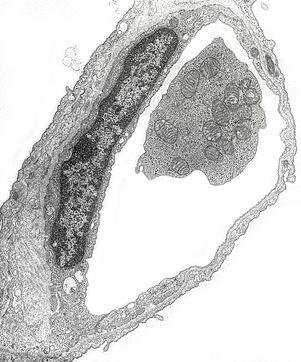
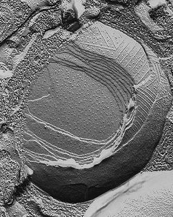
“This is a thin-section transmission electron micrograph (TEM) scan of a pulmonary capillary. My Tunica Intima image is created by a split through the inside of one of these cell membranes, exposing the amazing details on the inner surface.
The pulmonary capillary is one of the smallest blood vessels in the lung at the alveolar (deepest) level of the lung. The blood-air interface is very thin and enables oxygen exchange but it is actually composed of two cells each with two cell membranes.
I was interested in understanding the control of blood pressure by this surface and was able to localize an enzyme responsible for blood pressure control on the inside of these blood vessels, which led to the development of ACE Inhibitors, pharmaceutical drugs used primarily for the treatment of hypertension (elevated blood pressure) and congestive heart failure.”
— Una Ryan


Microscope as Art Medium
The electron microscope is a type of microscope that uses a beam of electrons to create an image of the specimen. It is capable of much higher magnifications and has a greater resolving power than a light microscope, allowing it to see infinitesimally smaller objects in finer detail. With their distinctive depth of field and incredible amount of detail, electron microscopes create stunning images of even the tiniest objects. They are large, expensive pieces of equipment, generally standing alone in a small, specially designed room and requiring trained personnel to operate them.
Modern electron microscopes produce electron micrographs in three ways using specialized digital cameras or frame grabbers to capture the image:
Freeze Fracture Electron Microscopy
Freeze fracture is unique among electron microscopic techniques in providing planar views of the internal organization of membranes. The technique consists of physically breaking apart (fracturing) a frozen biological sample; structural detail is then visualized by vacuum-deposition of platinum-carbon to make a replica for examination in the transmission electron microscope. Additional deep etching of ultra-rapidly frozen samples permits visualization of the surface structure of cells and their components.
Transmission Electron Microscopy (TEM)
The original form of electron microscope, the transmission electron microscope (TEM) uses a high voltage electron beam to create an image. When it emerges from the specimen, the electron beam carries information about the structure of the specimen that is magnified by the objective lens system of the microscope.
Scanning Electron Microscopy (SEM)
Unlike TEM, where electrons of the high voltage beam carry the image of the specimen, the electron beam of the scanning electron microscope (SEM) does not at any time carry a complete image of the specimen; it provide signals carrying information about the properties of the specimen surface, such as its topography and composition.
Wonders Within Our Living Cells
Life scientists such as Dr. Una Ryan engage in cellular and structural biology research to understand how cells function and respond to disease or genetic variations. Cellular biology explores individual cells and ways in which they are organized into organs and tissues. Structural biologists delve deep into sub-cellular components, organelles and macromolecular structures to unravel the fundamentals of life, which may consequently lead to solutions for treating fundamental diseases and disorders.
